Anthrax is a disease that is almost always associated
with
bioterrorism. Outside of this light though, many do not know just what it is or
where it comes from. Many people today think that Anthrax only recently came
about as it is portrayed in the media. This belief is not true and as we will
find, it has a history that spans many hundreds of years.
1250 BC- To truly understand Anthrax we will have to travel
back, some say, as far as the time when Moses brought the 10 plagues to Egypt.
According to the Center for Disease Control (Nov 2013), “ Anthrax is thought to
have originated in Egypt and Mesopotamia. Many scholars think that Moses’ time,
during the 10 plagues of Egypt, anthrax may have caused what was known as the
fifth plague, described as a sickness affecting horses, cattle, sheep, camels
and oxen” (p.1).
1700s- In fact, the CDC (2013) reports that, the first
clinically descriptive anthrax were actually reported in 1752 by “Maret in
1752… and Fournier in 1769” (p.2). It was reported that before this, anthrax
had only been described through historical accounts.
1800s-During the 1800s, doctors started to see the disease
of Anthrax but had not yet identified the pathogen. Their patients mainly
involved the workers of the clothing mills at the time and so the disease was
named “ wool sorters disease” (CDC 2013 pg.4). Then in the late 1800s, a man
named Robert Koch developed a set of postulates based upon Bacillus anthracis, which is the bacterium that causes anthrax. He
was able to study the rod shaped bacteria and found that part of the reason it
was so resilient was that the bacteria produces spores. Spores are a protein
coat that the bacteria produce when it is put under certain stress. Because of
this extra protein coat, the bacteria were able to survive long periods in very
extreme conditions. His work lead him to be able to culture the disease and
monitor the effects that it had on animals. The CDC explains that, “ [The]
Koch’s postulates… demonstrate a causal relationship between a specific microorganism
and a disease” (p.3). These postulates are an important part of immunology
today and helped to form the basis of today’s study of disease. Then, in 1881,
along came the father of modern immunology Louis Pasteur. Louis Pasteur was
able to take Koch’s work further by actually looking at the pathology of the
disease, or how the disease affected humans. He did this with the intent of
creating a Vaccine and so he did. He was able to create the first Animal
Anthrax vaccine in 1881 (CDC 2013). While this was shown to be a great step in
the cure for Anthrax, his vaccine only worked on animals. It wasn’t until the
1950s that the first human anthrax vaccine was created (CDC 2013). The vaccine
was given as a test to a group of goat hair mill workers. They were tracked
over a case of two years and it was determined that the vaccine was 92.5%
effective in preventing cutaneous anthrax (CDC 2013). The form of vaccine we
use today is based on an updated version from 1970.

Anthrax and Bioterrorism
Anthrax has been getting attention in recent years due to
its deadly onset and use as a bioweapon. The CDC has a full report estimating
Anthrax to be the most likely agent used in a bioterror operation. According to
the CDC (2014), “ If a bioterrorist attack were to happen, Bacillus anthracis, the bacteria that causes anthrax, would be one
of the biological agents most likely to be used. Biological agents are germs
that can sicken or kill people, livestock, or crops. Anthrax is one of the most
likely agents to be used because,
·
Anthrax spores are easily found in nature, can
be produced in a lab, and can last for a long time in the environment.
·
Anthrax makes a good weapon because it can be
released quietly and without anyone knowing. The microscopic spores could be
put into powders, sprays, food and water. Because they are so small you may not
be able to see, smell, or taste them.
·
Anthrax has been used as a weapon before.
Anthrax has been used as a weapon around the world for
nearly a century. In 2001, powdered anthrax spores were deliberately put into
letters that were mailed through the U.S. postal system. Twenty-two people,
including 12 mail handler, got anthrax, and five of these 22 people died”
(p.1).
Pathogenisis:
Anthrax is
caused by a bacteria called Bacillus anthracis. The disease is
found in three main forms; Inhalation Anthrax, Gastrointestinal Anthrax, and
Cutaneous Anthrax. Some important pathogenic factors of Bacillus anthracis include the fact that
it is a large (relatively 1.0 to 1.5 mcm x 3.0 to 5.0 mcm), gram-positive
bacillus (rod shaped) bacterium that is able to produce an endospore (Center
for Infectious Disease Research and Policy 2013). The bacterium is able to form
long chains of the vegetative form (non spore form) and also it is aerobic
(needs oxygen to survive). According to
the Center for Infectious Disease Research and Policy at the University of
Minnesota (2013), the bacteria is non-motile. This means that it is not able to
move on its own. This is a good thing since they are more confined to a location
and rest upon dependency of a vehicle or possibly a vector to transmit them to
a new host. The CIDRAP at the University of Minnesota (2013) lists some of the
most common virulence factors associated with the bacteria. First on its list
is LF or Lethal Factor.
Lethal factor (LF) is a zinc metalloprotease. LF combines with PA
to form lethal toxin. The PA is a Protective antigen and is a binding protein
that permits the entry of toxin into host cells through a process called
endocytosis. The PA forms the hole in the membrane of the host that allows the
LF to penetrate into the cytoplasm of the cell. The combination of the two is
called Lethal Toxin (CIDRAP 2013). LT is
thought to stimulate the over production of cytokines. Cytokines are
immunological components that help fight infection and cellular regulation from
macrophages. This factor will actually cause a lysis of the macrophages (CIDRAP
2013). The CIDRAP (2013) also illustrates the effectiveness of Lethal Toxin by
stating, “Lethal toxin has been shown to cause endothelial cell apoptosis and
endothelial barrier dysfunction, which may contribute to vascular destruction
(Kirby 2004, Warfel 2005). It has also been shown to reduce myocardial function
(Moayeri 2009, Sweeney 2010).” As if that wasn’t bad enough we also have an
additional toxin called the Edema toxin. The Edema toxin converts ATP to cAMP
in the intercellular compartment. This will lead to a hypotonic environment as
high levels of cAMP lead to impaired water homeostasis maintenance (CIDRAP 2013).

As stated earlier there are 3 main types of Anthrax;
Cutaneous Inhalational and Gastrointestinal.
Inhalational Anthrax:
As it maybe presumed, Inhalational Anthrax is considered the
most deadly. Inhalational Anthrax occurs in the following steps (CIDRAP 2013).
Firstly, the endospores are introduced into the body through the pathway of
inhalation. Because of their size they are able to reach the alveoli. Within
the alveoli the endospores are inactive, meaning that they are not able to
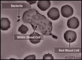
multiple or perform complex metabolic functions. At this stage they are then phagocytized
by macrophages as they body tries to quell the unknown invaders. The spores are
then taken to a regional lymph node in order to be destroyed and processed. It
is at this location that they are reanimated and take on their vegetative form.
It is at this point that they begin to multiply within the lymphatic system.
After the infection grows, pressure is placed on the mediastinum due to
inflammation and regional hemorrhagic lymphadenitis (Abramova 1993). This widening of the mediastinum is noted on
chest radiograph or enlarged lymph nodes can also be directly visualized on a
chest CT scan. If the lymphatic system becomes blocked then pulmonary edema may
occur. Pulmonary edema is a very dangerous condition in which there is a build
up fluid within the lungs. After some time, the bacteria may be able to enter
the bloodstream and cause septic shock and bacteremia. The stress placed on the
lungs and septic shock are the most common causes of death within this class.
The infectious dose of an organism refers to the amount of cells that is takes
to cause an infection within a person. According to the CIDRAP (2013), “ The
median infective dose… for inhalational anthrax is estimated at 8,000 to 50,000
spores (Franz 1997), although the minimum infective dose may be considerably
lower” (p.1).
Cutaneous
Anthrax:
Cutaneous
Anthrax occurs in the following stages. The initial presentation is similar to
that of inhalational Anthrax. The endospores are introduced through the skin
from a preexisting lesion or abrasion. In this case they are somewhat
opportunistic. Once the bacteria is present in the subcutaneous layer of the
skin the return to their vegetative form. Initial, early infections are characterized
by a localized necrosis with a soft-tissue or mucosal edema (Bischof 2007).
Because the wound will be easily visible quick antibiotic is enough to
eradicate the infection (CIDRAP 2013).
Gastro-Intestinal
Anthrax:
This form
of Anthrax is relatively rare and is caused by the ingestion of Anthrax spores.
Because it is so rare not as much information is available about this form as
the other forms of the disease. It is thought that not only is the disease
caused by ingestion of the spore form of the disease but also the vegetative
form (Inglesby 2002). There are two forms of gastrointestinal anthrax:
oropharyngeal and abdominal. According to the CIDRAP (2013), “In oropharyngeal
anthrax, the portal of entry is the oral or pharyngeal mucosa. A mucosal ulcer
occurs initially, followed by regional lymphadenopathy and localized edema. In
abdominal anthrax, the portal of entry often is the terminal ileum or cecum.
Intestinal lesions occur and are followed by regional lymphadenopathy. Edema of
the bowel wall and ascites (sometimes massive) may be present. Hematogenous
spread with resultant toxemia can occur”
(p.1).
Signs and
Symptoms:
Inhalation
anthrax presents with Flu-like symptoms. These include mild fever, fatigue and
muscle aches, which may last a few hours or days. As mentioned previously the
pathophysiology of the disease places a great pressure on the mediastinum in
the body, so it is to be expected that you should have mild chest discomfort,
shortness of breath, coughing up blood, painful swallowing and even nausea. In
the later stages you may have trouble breathing, present signs of shock or even
meningitis (Mayo clinic). It is
important to note that those most susceptible to the disease are those who work
with animal hides that come from the middle east and Africa. This is the most
common naturally occurring transmission of the disease.
Gastrointestinal
anthrax presents in some similar ways as inhalational anthrax, such are;
nausea, vomiting fever, sore throat and difficulty swallowing. Some additional
symptoms are; abdominal pain, headache, loss of appetite severe, bloody
diarrhea in the late stage and a swollen neck (Mayo Clinic).

Subcutaneous
anthrax will present as a raised, itchy red bump that appears to be an insect bite
and develops into a painless sore black centered lesion. The black center is a specific
sign of the anthrax disease as it is very distinct (Mayo Clinic).
Treatment
Options:
According to the CDC (2014), “Doctors have several options for
treating patients with anthrax, including antibiotics and antitoxin. Patients
with serious cases of anthrax will need to be hospitalized. They may require
aggressive treatment, such as continuous fluid drainage and help breathing
through mechanical ventilation.” (p.1). This is called a thoracocentesis and is used for the treatment of pleural effusions and pulmonary edema.
Antibiotics:
Any of the different types of
anthrax can be treated with different antibiotics. The use of certain
antibiotics depends on patient allergy but it is not uncommon to still see
penicillin used as an antibiotic to treat anthrax (CDC 2014).
Antitoxin:
As we went over earlier the main
virulence factor for the disease is its combined bacteriotoxins. To combat this
the CDC replies that there are different antitoxin treatments available. This
treatment needs to be done as soon as possible if an individual is suspected of
having anthrax especially inhalation anthrax (CDC 2014).
It is important to know that there is a vaccine available for both inhalation anthrax and cutaneous anthrax (CDC n.d). However, it is common to give this vaccine only to those individuals who are associated with high risk areas. The military is a wonderful example of a populous who is deployable to high risk areas. They will not even give it to the military members until they are about to breech a high risk area. The CDC reports that, "Currently, FDA has not approved the vaccine for use after exposure for anyone. However, if there were ever an anthrax emergency, people who are exposed might be given anthrax vaccine to help prevent disease. This would be allowed under a special protocol for use of the vaccine in emergencies" (p.1). The vaccine consists of 5 shots over 18 months and an annual booster. The CDC seems to lean towards not giving the vaccine to everyone because it may carry a higher risk of allergic reactions though, they are not clear on exactly why this is. Allergic reactions can happen to any treatment and they had said previously that the vaccine did not contain an active form of B. anthracis. I could only see the effective treatment plan being increased with additional public knowledge available about why regular population can not get vaccinated. And if after that data does become available that they be able to disperse these vaccinations to those who are not subject to the contraindications.
References
Bischof, T.,
Hahn, B., & Sohnle, P. (2007). Characteristics of Spore
Germination in a Mouse Model of Cutaneous Anthrax. Journal of
Infectious Diseases, 195(6), 888-894. doi:10.1086/511824
Center for
Disease Control and Prevention (2014, November). Who Is At Risk |
Anthrax | CDC. Retrieved June 15, 2014, from
http://www.cdc.gov/anthrax/risk/index.html
Davison, S.,
Couture-Tosi, E., Candela, T., Mock, M., & Fouet, A.
(2005). Identification of the Bacillus anthracis Phage Receptor. Journal
of Bacteriology, 187(19), 6742-6749.
doi:10.1128/JB.187.19.6742-6749.2005
Klee, S. R.,
Brzuszkiewicz, E. B., Nattermann, H., Brüggemann, H.,
Dupke, S., Wollherr, A., . . . Liesegang, H. (2010). The Genome
of a Bacillus Isolate Causing Anthrax in Chimpanzees Combines Chromosomal
Properties of B. cereus with B. anthracis Virulence Plasmids. PLOS One, 5(7).
doi:10.1371/journal.pone.0010986.t002
New York State, Dept of Health (2011, October
11). Anthrax (malignant edema, woolsorters' disease).
Retrieved June 15, 2014, from
http://www.health.ny.gov/diseases/communicable/anthrax/fact_sheet.htm
University of
Minnesota (2013, May 1). Anthrax | CIDRAP. Retrieved June 15,
2014, from http://www.cidrap.umn.edu/infectious-disease-topics/anthrax
References for Media
(In order of appearance)
http://www.cdc.gov/anthrax/history/index.html
http://www.cdc.gov/anthrax/bioterrorism/threat.html
http://giphy.com/gifs/A30hVv6SbTNFm
http://giphy.com/gifs/growth-bacteria-bacillus-kwSe0Dw7rIXFS
http://en.wikipedia.org/wiki/Anthrax